![]()
Medical
Design Online - June 2009
Industry News
'Dyeing' to figure out how the brain works
Voltage-sensitive dye and optical recording techniques are giving neuroscientists at Georgetown University's Department of Physiology and Biophysics a new means for figuring out how the brain works.
 Professor Jian-Young Wu and his colleagues are conducting research on waves of neuronal activity. They visualize wave-like patterns in the brain cortex using a new method called voltage-sensitive dye imaging. The dye binds to the membrane of neurons and changes color when electrical potential changes on the membrane of active neurons. The neuronal sample is derived from slices of rat neocortex.
The neocortex is the outer layer of the cerebral hemispheres in the brain of mammals. Made up of six layers, it is involved in higher functions such as sensory perception, generation of motor commands and, in humans, language.
The neurons of the neocortex are arranged in vertical structures called neocortical columns. These are patches of the neocortex with a diameter of about 0.5 mm and a depth of 2 mm. Each column typically responds to a sensory stimulus representing a certain body part, or region of sound or vision, hi the human neocortex, there are believed to be about a half-million of these columns, each of which contains approximately 60,000 neurons.
The neocortex can be viewed as a huge web, consisting of billions to trillions of neurons and hundreds of trillions of interconnections. While individual neurons are too simple to have intelligence, the collective behavior of the billions of interneuronal interactions occurring each second can be highly intelligent.
Traditionally, scientists have studied brain activity by placing a few electrodes in the brain and measuring the electrical signals of the neurons close to the electrodes. This method is workable for understanding the function of the cortex and interactions between individual neurons, but it is not suitable for studying the emerging properties of the nervous system.
"It is like viewing a few pixels on a television screen and trying to figure out the story," explains Wu. "Now, with optical methods and voltage-sensitive dyes, we can visualize the activation in a large area of the neocortex when the brain is processing sensory information, similar to watching the whole television screen."
"Voltage-sensitive dye is a compound that stains neuronal membrane and changes its color when the neuron is excited," continues Wu. "This allows us to visualize population neuronal activity dynamically in the cortex. We study how individual neurons in the neocortex interact to generate population neuronal activities that underlie sensory and motor processing functions. Population activities are composed of the coordinated activity of billions of neurons. Currently, we study how oscillations and propagating waves can be generated by small ensembles of neocortical neurons."
Viewing the spatiotemporal patterns of neuronal population in the cortex is markedly different from recording individual neurons. Here the cortical activity is viewed as "population activity," which can be more complex than the linear addition of an individual neuron's activity. Voltage-sensitive dye and optical recording techniques give the neuroscientist new tools for figuring out how brain cortex works.
Negative-stiffness mechanism vibration isolation systems from Minus K Technology enabled the lab to get vibration isolation down to a level of 1 Hz.
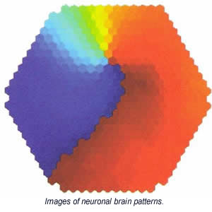
Wu's imaging team has uncovered spiraling wave patterns resembling little hurricanes in the brain. He believes that this hurricane-like spiral pattern is an emergent behavior of the network.
"A metrological hurricane is an emergent behavior of a large volume of air molecules," says Wu. "If you were to dissect a hurricane into individual air molecules you would not find any special process that generates a hurricane. Similarly, in the nervous system, spiral waves are an emergent process of the neuronal population and there might be no special cellular process attributed to spirals."
However, like a hurricane, spiral waves can be a powerful force. Their power is seen when it comes to organizing the activity of a neuronal population. Spirals generated in a small area can send out a powerful storm that invades large, normal brain areas and starts a seizure attack. This hypothesis means that epilepsy could be viewed not just as "mis-wiring" in the brain, but as an abnormal wave pattern that invades normal tissue.
Similarly, during cardiac fibrillation, spiral waves form in the heart emitting rotating and scroll waves in two and three dimensions. As a leading life-threatening situation, these rotating waves can kill the patient instantly as the pumping function of the heart is disrupted by the 5 to l0 Hz rotations, which drives chaotic and abnormally rapid cardiac contractions.
Wu believes that propagating waves are a basic pattern of cortical neuronal activity, and that these wave patterns may play an important role in initiating and organizing brain activity involving millions to billions of neurons. Studying the spatiotemporal patterns of neuronal population activity may provide insight into normal brain functions and pathological disorders. This research has the potential to help scientists understand abnormal waves that are generated in the brains of patients with epilepsy.
Since voltage-sensitive dye signals are small - a change of 0.1 to 1.0 percent of the illumination intensity - Wu's team uses a high-dynamic-range camera, photodiode array to detect the voltage-sensitive dye signals of the cortical activity. The photodiode array can resolve extremely small changes in light, usually one part, of ten thousands. (Human eyes and ordinary digital cameras register light changes of one part to a hundred). Detecting such small signals requires an extreme isolation of vibration. The lab had to contend with low frequency vibrations from air conditioning equipment, people walking, and wind blowing against the building. Vibrations as low as 1 Hz were inhibiting the integrity of the images and data.
"At first, we used high quality air tables, but they were not adequate for isolating low frequency vibrations," says Wu. "We tried putting a second air table on top of the first one, but that still did not give us the isolation we needed. Then we tried an active, electronic system, but we were still spending much time fighting with floor vibrations. We were dealing with an unresolved vibration problem for many years."
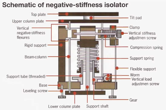
The Georgetown lab eventually tested, and settled upon negative-stiffness mechanism vibration isolation systems from Minus K Technology, Inglewood, CA, (www.minusk.com) which enabled the lab to get vibration isolation down to a level of 1 Hz. This effectively cancelled out any vibration noise difficulties that were inhibiting image and data readings.
Negative-stiffness mechanism (NSM) isolators have the flexibility of custom tailoring resonant frequencies vertically and horizontally. They employ a completely mechanical concept in low-frequency vibration isolation. Vertical-motion isolation is provided by a stiff spring that supports a weight load, combined with a NSM. The net vertical stiffness is made very low without affecting the static load-supporting capability of the spring.
Beam-columns connected in series with the vertical-motion isolator provide horizontal-motion isolation. The horizontal stiffness of the beam-columns is reduced by the "beam-column" effect. A beam-column behaves as a spring combined with a NSM. The result is a compact passive isolator capable of low vertical and horizontal natural frequencies and high internal structural frequencies.
Transmissibility with negative stiffness is substantially improved over air systems, which can make vibration isolation problems worse since they have a resonant frequency that can match that of floor vibrations. Transmissibility is a measure of the vibrations that transmit through the isolator relative to the input vibrations. The NSM isolators, when adjusted to 0.5 Hz, achieve 93 percent isolation efficiency at 2 Hz; 99 percent at 5 Hz; and 99.7 percent at l0 Hz.
NSM transmissibility is also improved over active systems. Active systems run on electricity and can be negatively influenced by problems of electronic dysfunction and power modulations, which can interrupt scanning. They also have a limited dynamic range that is easy to exceed. This causes the isolator to go into positive feedback and generate noise underneath the equipment. Although active isolation systems have fundamentally no resonance, their transmissibility does not roll off as fast as negative-stiffness isolators.
Within the past 10 years, Wu's team has documented a variety of waveforms (e.g., plane wave and spirals) in brain slices during artificially induced oscillations. Using neocortical slices and mathematic models they are studying the initiation of the waves and the factors that control their propagating direction and velocity.
The negative-stiffness vibration isolation system are also used by the lab in the development of optical imaging techniques such as imaging propagating waves in vivo, within intact brains. "This is technically difficult," says Wu. "Other imaging methods (MRI, PET or MEG) provide inadequate spatiotemporal resolution. Large scale neuronal activity is a hallmark of a living brain. We hope to visualize the waves in the cortex in vivo during sensory processes and in animals while behavioral and cognitive tasks are performed."